A research reactor is primarily a source of high-intensity neutrons. To a lesser extent, gamma radiation is also used, either accompanying nuclear reactions involving neutrons or emitted during radioactive decay e.g. from spent fuel.
The reactor itself and the processes taking place in it are also sometimes the subject of research, especially when nuclear safety and radiation protection issues are involved. The reactor can also be used for teaching in reactor physics and technology.
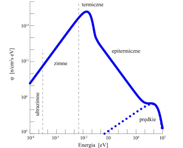
Neutrons emitted during fissions have energies on the order of a few megaelectron volts and are referred to as fast neutrons (Figure 1). As a result of collisions with the nuclei of the medium, mainly the moderator, a region of epithermal neutrons appears in the neutron spectrum with a characteristic 1/E-type energy dependence. After reaching energies of the order of several meV, a state of thermodynamic equilibrium between the neutron energies and the thermal vibrational energies of the medium is established. Hence the name: thermal neutrons. In this region, the neutron energy spectrum is characterised by a Maxwell distribution. Due to the use for physical research in the low energy region, the cold and ultra-cold neutron regions are still separated.
Neutrons from all energy ranges find their application. Fast neutrons in so-called threshold reactions, i.e. nuclear reactions that occur with neutrons of a certain minimum threshold energy. In the neutron energy region above 0.1 eV, resonant reactions occur whose active cross sections have a characteristic resonant character. Thermal neutrons are most commonly used in radiation capture reactions; the active cross sections of these reactions are usually of the characteristic 1/v type.
The de Broglie wavelengths of thermal neutrons are comparable to the interatomic distances in a solid and hence the widespread use of these neutrons for research in condensed matter physics.

To produce radioisotopes in the MARIA reactor, dozens of so-called isotope channels are used, characterised by different neutron intensities and energy spectra. The target materials from which the radioisotopes are produced are generally irradiated in aluminium containers.
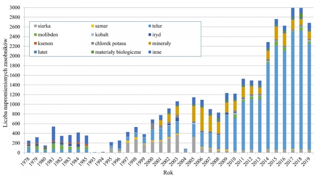
Due to the annual activities obtained, the list of radioisotopes produced in the MARIA reactor opens with: 131I, 35S, 32P, 153Sm, 169Yb, 60Co and 177Lu. Most of the radioisotopes coming from the MARIA reactor are isotopes used in nuclear medicine.
The production of radioisotopes, expressed in terms of the number of containers with target materials irradiated in successive years of MARIA reactor operation, is shown in the figure.
A particularly important radioisotope, used in 80% of nuclear medicine procedures, is technetium-99m. This isotope is a decay product of 99Mo (this decay occurs in so-called technetium generators). Molybdenum-99 can be produced by activating the natural isotope 98Mo, but the most efficient source of 99Mo is the fission process of uranium-235.
In the MARIA reactor, uranium targets for 99Mo production are irradiated in specially adapted fuel channels. Thanks to the efficient cooling system of the fuel channel cooling circuit, high fission densities and thus high 99Mo activities can be achieved in the uranium plates. The duration of irradiation of the plates is about 140 hours, allowing 7500 Ci of 99Mo to be obtained from 40 grams of uranium-235 at the end of activation. After irradiation and a cooling period of several hours, the highly active uranium plates are transported to the reprocessing plant (Petten, the Netherlands), where the 99Mo isotope is extracted.
About 10% of the world's demand for this isotope is obtained from the uranium targets irradiated at MARIA; in the record year of 2014, when it was necessary to replace other reactors temporarily shut down due to maintenance, it was as much as 20%.

The interaction of neutrons with matter leads not only to the formation of radioactive isotopes, but often to changes in the properties of materials. This is the case, for example, with the irradiation by fast neutrons of certain high-temperature superconductors, where a significant increase in critical currents is observed as a result of such irradiation.
Another example of changing the properties of materials is the colouring of topaz minerals. Naturally occurring topaz minerals are colourless. Irradiating such minerals with fast neutrons changes their colour to blue. In the MARIA reactor, the colouring of topaz minerals is carried out in specially adapted isotope channels, in a graphite reflector. The installations use thermal neutron shielding to minimise unwanted activation of the minerals.
Reactor neutrons are also used in the neutron doping of silicon (NTD) process. The physical basis of this process is the 30Si (n,) 31Si 31P reaction. The resulting stable phosphorus isotope is an n-type dopant in silicon. The semiconductor properties ( specific resistance) of silicon are determined by the level of phosphorus doping, i.e. the 31P concentration. This in turn depends on the fluence (dose) of thermal neutrons received by the silicon in the reactor.
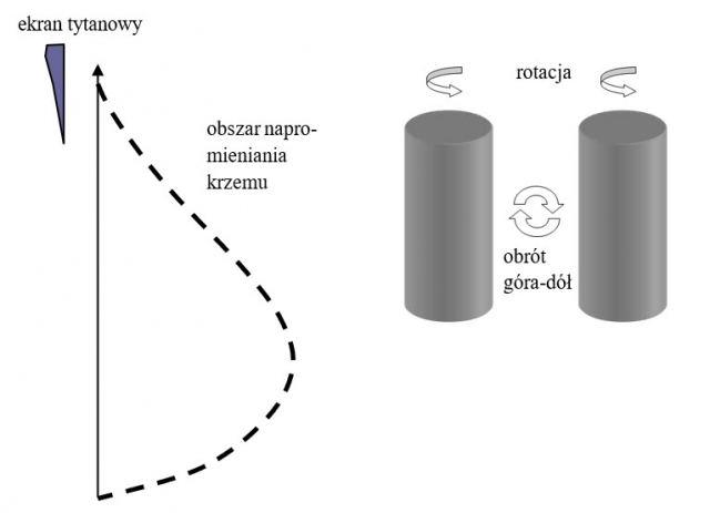
The key quality parameter of NTD technology is the homogeneity of the neutron field in the silicon, because only then can a homogeneous level of doping, superior to classical doping methods, be achieved. In the MARIA reactor, silicon samples of considerable size (cylinders 50 cm high and up to 6 in diameter) are irradiated in a specially adapted channel. The figure shows schematically the silicon irradiation area in the silicon installation and how the axial and radial uniformity of the neutron dose is ensured. In addition to the continuous rotation of the sample during irradiation (radial homogeneity), the silicon cylinder is inverted up and down in the middle of the irradiation cycle (axial homogeneity).

(historical material; the experimental hall is currently being upgraded, where new research equipment will be installed)
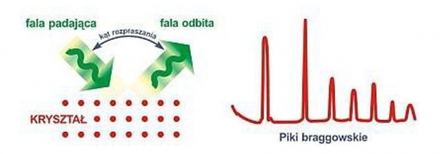
From the MARIA reactor, neutron beams are routed out, mostly used for condensed matter research. Five of the 6 horizontal channels currently available are equipped with instruments that take advantage of neutron wave scattering effects (diffractometers, triaxial spectrometers). The physical basis of neutron scattering on crystalline structures is illustrated by the diagram in Figure 1.

The ultra-low-angle diffractometer allows, among other things, the study of the size of magnetic domains in amorphous, nanocrystalline and polycrystalline materials, as well as the influence of external factors such as temperature, mechanical stress and magnetic field on the size of these domains. The low-angle neutron scattering diffractometer is designed to study the fine (1 nm 30 nm) inhomogeneities present in materials, while the high-resolution diffractometer is used to study the quality of metal monocrystals, among other things. A diffractometer designed for atomic and magnetic ordering studies can be used as a polarised neutron diffractometer.
Triaxial thermal neutron spectrometers are mainly used to study the collective motions of atoms (phonons) and magnetic moments (magnons) by inelastic (coherent) neutron scattering. They are also used to study electron energy levels in ions forming a solid by inelastic (incoherent) neutron scattering.
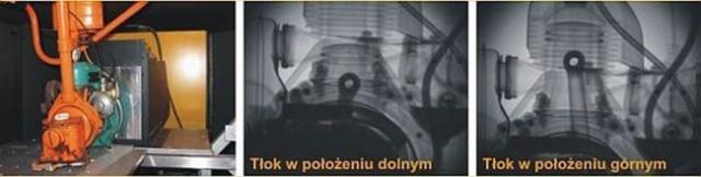
A neutron and gamma radiography station is installed at one of the horizontal channels (H8). Radiography is a method of obtaining images of the interiors of objects that are opaque to visible light. An object that completely absorbs light radiation can be almost transparent to other types of radiation, such as X-rays, gamma or neutron radiation. When image capture makes it possible to observe the movement of the object under examination, one speaks of dynamic radiography. The dynamic neutron and gamma radiography facility at the MARIA reactor uses the neutron and gamma radiation produced in the reactor. Particularly a great deal of information is obtained through the use of neutrons. At present, the applications of neutron radiography focus on the study of static objects (internal structure of technical equipment, detection of defects and heterogeneity of materials) and on the study of processes such as water migration in porous materials, fluid transport in filter beds, development of the root system of plants, transport of crude oil in geological deposits, etc.
")
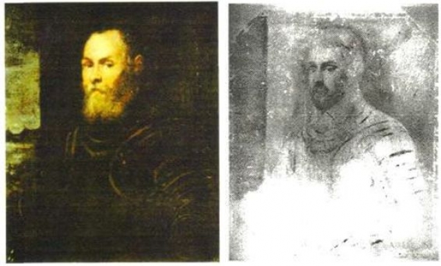
The neutron beam from the horizontal channel can also be used to study paintings using the technique of neutron autoradiography. Such a technique was used in the MARIA reactor to study paintings by Venetian painting masters of the 14th to 18th century from the collection of the National Museum in Warsaw, presented as part of the exhibition 'Serenissima - the light of Venice' in 2000.
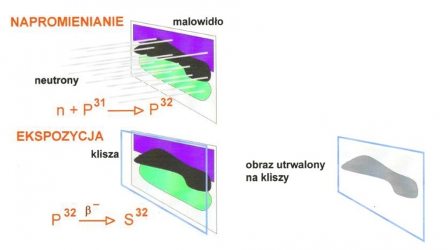
The autoradiography technique involves irradiating the paintings with thermal neutrons and triggering a radiation capture reaction in some of the isotopes that make up the painting layers, such as 31P (n, γ) 32P. At the end of the neutron irradiation process, decay (generally beta decay) of the resulting radioactive isotopes takes place, e.g. 32P→32S. If the image is covered with β- X-ray film at this time, the emitted particles cause a blackening of the film. The pattern formed on the film reflects the distribution of the isotope - emitter of the beta particles in the painting layer. This applies not only to the surface of the image, but also to the deeper structures of the paint layer. The image of the blackening of the film is characteristic of the pigment used and allows analysis of the deeper painting layers. Fig. NCBJ-4.6 shows schematically the autoradiography technique of the paintings.

A spectacular example of the use of this technique is the autoradiography of the painting 'Portrait of the Venetian Admiral' by Jacopo Tintoretto. Figure 2 shows the original and one of the autoradiograms, indicating the existence of another portrait under the actual painting.


